 English
 Spanish
 French
 German
 Italian
 Chinese (Simplified)
 Japanese
 Korean
 Arabic
 Portuguese
 Get A Quote
Showroom

(10)
These durable and trusted Laboratory Machines Spare Parts And Kits are available in different sizes to suit the varied demands of models of lab machines.

(8)
Laboratory Chemicals are available in precise composition for ensuring to provide exact results with high level of safety. They are provided in bulk in high-quality packaging materials.

(10)
UV Visible Spectrophotometer is developed in durable designs for measuring light intensity/absorbency of a material's reflection or transmission at a specific wavelength.

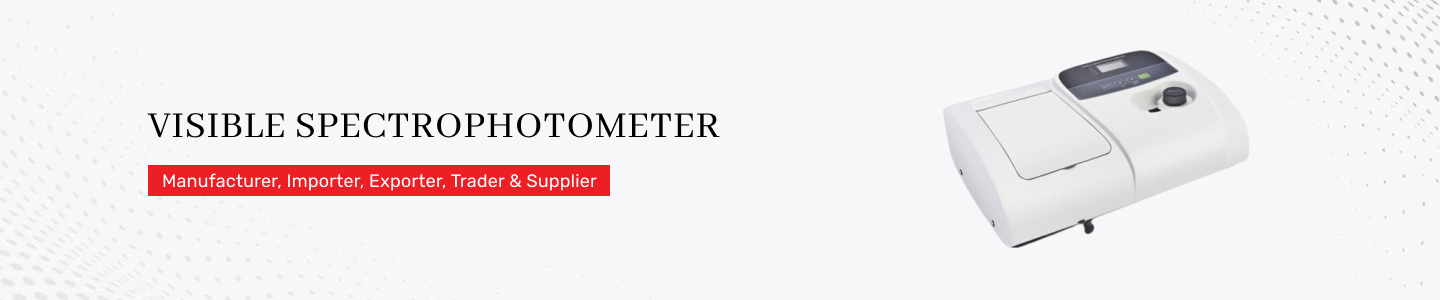
(2)
The maximum scan rate of this Visible Spectrophotometer is 24,000 nm per minute. It is controlled by the advanced software with its modular design.

(2)
FTIR Spectrophotometer is ideal to be used if the spectral region is in the infrared or high spectral resolution is necessary. This is designed in durable structure to provide the best and accurate results.

(22)
Atomic Absorption Spectrometer is designed to be used in different sectors such as environmental testing, petroleum and chemical production, metal analysis, semiconductor manufacturing, pharmaceuticals, etc.

(2)
Fused silica glass made HID Xenon Lamps have wide application in pharmaceutical, semiconductor and research and analysis arena. These lamps are reckoned for their high luminous efficacy and stable performance.

(2)
Autosampler Vials, Caps And Closures are used for secure storage of liquid injection. Made of non toxic grade raw materials, these cost effective pharmaceutical containers and accessories are offered in sterilized form.

(6)
Hand Held Refractometers are reckoned for their precise measurement method. Featured with automatic calibration mechanism, these instruments are preferred for their ease of gripping, ergonomic design, utilization of natural light and consistent operation.

(22)
Laboratory Platinum Ware is known for its high temperature proof design, oxidation and rust protected surface. Offered in different specifications, this laboratory product is deformation proof and it has unique tensile strength.

(38)
Equipped with RS-232 communication software, this PH Meter is instrumental in assessing alkalinity level of any solution. High precision level, automatic calibration function and error detection arrangement are its key factors.

(4)
Conductivity Meter is used to measure conductivity level, TDS rate, temperature and salinity of water. Automatic operation, easy to read display screen and application of RS-232 communication software are its key features.

(3)
Dissolved Oxygen Meter is used to assess amount of oxygen present in waste water and aquarium. This system uses USB interface to transfer measured data to computer. Accurate operation and ergonomic design are its key features.

(15)
You always need a good instrument in order to do color analysis of products, like chemicals, textiles, paper and more. Our company can help businesses find the right color measurement instrument.

(1)
Digital ultrasonic cleaner is required to remove dust, dirt, fingertips and more from the lab tools and instruments. This is a high efficiency cleaner that can quickly clean instruments and tools in an effective way.

(2)
Laboratory Blenders are occasionally preferred over a typical large-scale production-size device because the pharmaceutical, food, and chemical industries frequently require to mix a tiny quantity of product for some specific manufacturing process. They are very effective and safe to use.

(9)
Laboratory Microscopes are used to magnify blood samples so that doctors can see the malaria parasites attacking the red blood cells. They aid in the creation of the extremely small electrical circuits found in Silicon microchips. They require very low maintenance and replacement costs.

(3)
Hollow Cathode Lamp improves lamp performance while also cutting down on warm-up time. This is made feasible by stringent calibration and maintenance procedures for laboratory instruments and volumetric glassware. This lamp is tested under various parameters to ensure its high quality.

(3)
Digital Anemometer can measure atmospheric effects such as wind speed, temperature, and humidity. This uses changes in a fluid's physical properties or its impact on a mechanical device put into the flow to determine the air velocity. This is safe to use.

(77)
HPLC Columns can interact with non-polar molecules in the liquid phase due to their high polarity. Worldwide, clinical diagnostics laboratories, industries, environmental, and forensic testing laboratories employ our HPLC and semi/prep columns. A computer will examine the components once they have been separated using column chromatography.

(3)
Reference Standards from novel pharmacological compounds intended for assays. They are deemed to be the best approach for classifying participants as having or not having a target condition in a study of diagnostic test accuracy. They are great to use.

(3)
Rheodyne Injectors and Spares are precision chromatography accessories used in HPLC systems for accurate sample injection and flow control. Widely utilized in pharmaceutical, chemical, biotechnology, and analytical laboratories, these components support reliable chromatographic performance, reduced downtime, and efficient instrument maintenance in routine analytical operations.

(2)
Sound Level Meters are precision instruments designed to measure noise intensity and sound pressure levels in industrial, environmental, and laboratory settings. Widely used in manufacturing plants, construction sites, automotive industries, and occupational safety monitoring, these devices help maintain regulatory compliance, workplace safety, and acoustic analysis. Advanced models support data logging, frequency analysis, and real-time environmental noise assessment.

(5)
Digital Stroboscope allows for non-contact examination and observation of moving parts. The top and bottom handles that are supplied help with transportation and usage versatility. This is tested under various parameters to ensure its high quality and effectiveness.

(13)
Filtration Products include membrane filters, filtration assemblies, and consumables used for separating particles, microbes, and impurities from liquids and gases. Commonly applied in pharmaceuticals, biotechnology, food processing, water treatment, and chemical laboratories, these products ensure sample purity, contamination control, and process efficiency. They are essential for quality assurance, analytical testing, and sterile laboratory operations.

(24)
Brookfield Viscometers are advanced viscosity measurement instruments used to determine the flow characteristics and consistency of liquids, semi-solids, and pastes. Widely utilized in pharmaceuticals, cosmetics, paints, food processing, petrochemicals, and polymer industries, these systems support product quality control, formulation development, and rheological studies. They provide accurate, repeatable viscosity data for research and industrial applications.

(3)
Brookfield Viscosity Standards are developed for the precise calibration of viscometers and rheometers. Compared to oil viscosity norms, silicone fluids offer good temperature stability and are less temperature sensitive. They are safe to use.

(11)
Electrophoresis Systems are laboratory instruments designed for the separation and analysis of DNA, RNA, and proteins using an electric field. Commonly used in biotechnology, molecular biology, pharmaceutical research, and clinical diagnostics, these systems support genetic analysis, protein characterization, and forensic applications. They provide reliable sample separation, high-resolution analysis, and efficient laboratory workflow.

(8)
Chemicals have no effect on the Syringe Filter Holders, and they don't contain any trace components that might leach into the liquid being filtered. The filter holder is helpful for ultra-cleaning HPLC samples, solvents, tissue culture media, and other items.

(5)
Numerous scientific and industrial uses exist for Vacuum Pump. In addition, attractive vacuum coatings on metal, glass, and plastics, energy-saving and durable glass, ophthalmic coatings, and hard coatings require a pump. This is very effective to use.

(1)
Nucleic Acid Electrophoresis Cells are specialized units used for separating DNA and RNA samples in agarose or polyacrylamide gels. Widely applied in molecular biology laboratories, biotechnology research, academic institutions, and diagnostics, these cells support genetic testing, PCR analysis, cloning, and sequencing applications. Their durable construction and efficient design ensure accurate migration and reproducible experimental results.

(1)
Gel Documentation Systems are imaging platforms used to capture, analyze, and document electrophoresis gels and blotting membranes. Extensively used in biotechnology, life sciences, pharmaceuticals, and academic research, these systems enable DNA, RNA, and protein visualization with high sensitivity. They support image analysis, quantitative measurements, and digital record management for molecular biology applications.

(1)
UV Transilluminators are laboratory devices used to visualize nucleic acids and proteins stained with fluorescent dyes during electrophoresis procedures. Commonly used in biotechnology, genetics, microbiology, and pharmaceutical research laboratories, these systems provide uniform UV illumination for accurate gel analysis. They are essential for DNA band detection, cloning studies, and molecular diagnostics applications.

(1)
PCR Genemate Series instruments are advanced thermal cyclers designed for DNA amplification and molecular diagnostics applications. Widely used in biotechnology, genomics, healthcare, food testing, and forensic laboratories, these systems support gene expression studies, pathogen detection, and research analysis. Their precise temperature control and user-friendly operation ensure reliable, high-throughput PCR performance.

(3)
FTIR and IR Sample Cards are accessories used for preparing and analyzing samples in Fourier Transform Infrared spectroscopy applications. Commonly applied in chemical analysis, pharmaceuticals, polymers, petrochemicals, and academic research, these cards provide efficient sample handling and accurate infrared spectral measurements. They support material identification, quality control, and analytical research processes.

(5)
IR Crystal Optics are precision optical components designed for infrared spectroscopy and analytical measurement applications. Used extensively in FTIR systems, pharmaceutical laboratories, petrochemical analysis, and material science research, these optics ensure high transmission efficiency and spectral accuracy. They are critical for ATR analysis, chemical characterization, and advanced infrared testing procedures.

(6)
Sealed Liquid Spectrophotometer Cells are specialized cuvettes designed for safe and contamination-free optical measurements of volatile or sensitive liquid samples. Widely used in pharmaceutical, chemical, environmental, and research laboratories, these cells support UV/VIS spectroscopy applications. Their leak-proof construction ensures accurate absorbance analysis, sample integrity, and reliable analytical performance.

(7)
Demountable Liquid Cells are reusable spectroscopy accessories designed for liquid sample analysis with adjustable path lengths. Commonly used in FTIR, infrared spectroscopy, chemical analysis, and pharmaceutical laboratories, these cells enable easy cleaning, flexible sample handling, and accurate spectral measurements. They are ideal for research, quality control, and analytical testing applications.

(5)
Flow Temperature Controlled and Pressure Cells are advanced analytical accessories used for studying samples under controlled temperature and pressure conditions. Extensively utilized in petrochemical, polymer, pharmaceutical, and material research industries, these cells support real-time spectroscopic analysis and process monitoring. They ensure precise environmental control for accurate characterization and industrial research applications.

(8)
Gas Cells are specialized spectroscopy accessories designed for the analysis of gaseous samples in infrared and UV/VIS systems. Widely used in environmental monitoring, chemical processing, petrochemical industries, and research laboratories, these cells support gas identification, emission analysis, and process control. Their durable design ensures accurate gas sampling and reliable analytical performance.

(12)
Infrared Accessories include sample holders, ATR crystals, windows, cells, and optical components used with FTIR and infrared spectroscopy systems. Commonly applied in pharmaceuticals, chemicals, polymers, environmental testing, and research laboratories, these accessories improve analytical flexibility, sample handling, and measurement accuracy. They support advanced material characterization and routine spectroscopic analysis.

(4)
Reconditioning services for ATR prisms, crystal windows, and spectroscopy cells restore damaged or worn analytical components for optimal performance. Widely required in pharmaceutical, chemical, and research laboratories, these services improve spectral accuracy, extend equipment life, and reduce replacement costs. They ensure reliable FTIR analysis and efficient laboratory operations.

(4)
Intrinsic Dissolution Apparatus are laboratory systems used to study the dissolution rate of pharmaceutical compounds under controlled conditions. Commonly utilized in pharmaceutical research, formulation development, and quality control laboratories, these instruments support drug solubility analysis, bioavailability studies, and regulatory compliance testing. They provide accurate dissolution profiling for drug development applications.

(13)
Solid Sampling and KBr Presses are laboratory devices used for preparing solid samples into pellets for FTIR spectroscopy analysis. Widely applied in pharmaceutical, chemical, mineral, and polymer industries, these systems ensure uniform sample preparation and accurate spectral measurements. They support analytical research, material characterization, and routine quality control testing.

(2)
Laboratory Grinding Mills are sample preparation instruments designed for grinding, pulverizing, and homogenizing solid materials. Commonly used in mining, pharmaceuticals, agriculture, food processing, and research laboratories, these mills support particle size reduction and analytical sample preparation. They improve consistency, accuracy, and efficiency in laboratory testing applications.

(3)
Internal Reflection Accessories are analytical components used in infrared spectroscopy for ATR and reflection-based sample analysis. Extensively utilized in pharmaceuticals, polymers, coatings, and chemical industries, these accessories enable rapid, non-destructive testing of solids, liquids, and films. They improve spectral accuracy, sample convenience, and analytical versatility.

(1)
Temperature Controllers are precision instruments designed to regulate and maintain stable temperatures in laboratory and industrial processes. Commonly used in pharmaceuticals, biotechnology, chemical manufacturing, and research facilities, these systems support incubation, reaction monitoring, and process automation. They ensure accurate thermal management, improved efficiency, and reliable experimental results.

(11)
UV/VIS Spectrophotometer Cells and Cuvettes are precision optical containers used for absorbance and transmission measurements in spectroscopy applications. Commonly utilized in pharmaceutical, chemical, food, environmental, and academic laboratories, these accessories ensure accurate sample analysis. They support quantitative testing, quality control, and research in analytical chemistry.

(1)
Stability Chambers are environmental testing systems designed to simulate controlled temperature and humidity conditions for product stability studies. Widely used in pharmaceuticals, cosmetics, food, and biotechnology industries, these chambers support shelf-life testing, accelerated aging, and regulatory compliance. They ensure reliable environmental control for research and quality assurance applications.

(4)
Digital Colony Counters are microbiology instruments used for accurately counting bacterial and fungal colonies on culture plates. Commonly used in pharmaceutical, food, water testing, healthcare, and research laboratories, these systems improve counting accuracy, reduce manual errors, and increase productivity. They support microbial analysis, quality control, and laboratory documentation.

(13)
Weighing Balances are precision laboratory instruments designed for accurate measurement of mass in analytical and industrial applications. Extensively used in pharmaceuticals, chemicals, food processing, jewelry, and research laboratories, these balances support formulation, testing, and quality assurance processes. They offer high sensitivity, reliability, and compliance with laboratory standards.

(4)
Flame Photometers are analytical instruments used for determining the concentration of metal ions such as sodium, potassium, and calcium in samples. Widely applied in clinical diagnostics, agriculture, food analysis, pharmaceuticals, and water testing laboratories, these systems provide rapid elemental analysis, quality control, and routine chemical testing.

(1)
Microscope Cameras are imaging devices designed for capturing high-resolution images and videos through microscopes. Commonly used in life sciences, pathology, material science, education, and industrial inspection, these cameras support documentation, image analysis, and research applications. They enhance visualization, digital reporting, and collaborative laboratory studies.

(1)
Vortex Shakers are compact laboratory mixers designed for rapid mixing of liquids in tubes, vials, and containers. Widely used in biotechnology, pharmaceuticals, microbiology, and chemical laboratories, these devices ensure efficient sample homogenization and preparation. They support routine laboratory workflows, diagnostic testing, and research applications with reliable mixing performance.

(4)
ROCKER Lab Instruments include vacuum filtration systems, pumps, and laboratory equipment designed for analytical and microbiological applications. Commonly used in pharmaceutical, environmental, food, and academic laboratories, these instruments support filtration, sample preparation, and contamination control. They are known for reliable performance, durability, and efficient laboratory operations.

(1)
Rotary Mixers are laboratory devices designed for gentle and continuous mixing of samples in tubes, bottles, and containers. Widely used in biotechnology, pharmaceuticals, clinical diagnostics, and research laboratories, these systems support blood sample preparation, chemical mixing, and biological assays. They provide uniform agitation and reliable laboratory performance.

(1)
Vacuum Desiccators are sealed laboratory containers used for moisture-free storage and drying of sensitive materials under vacuum conditions. Commonly applied in chemical, pharmaceutical, electronics, and research laboratories, these systems protect hygroscopic samples from humidity and contamination. They support sample preservation, drying processes, and analytical testing.

(3)
Micro Centrifuges are compact laboratory instruments used for rapid separation of small-volume liquid samples through centrifugal force. Commonly applied in molecular biology, clinical diagnostics, biotechnology, and pharmaceutical research, these devices support DNA extraction, sample preparation, and protein analysis. They provide efficient, high-speed separation with reliable performance.

(6)
Rotary Evaporators are laboratory systems designed for efficient solvent evaporation, concentration, and distillation under reduced pressure. Extensively used in chemical, pharmaceutical, food, and research laboratories, these instruments support sample purification, extraction, and solvent recovery. They ensure precise temperature control, faster processing, and reliable laboratory performance.

(1)
Cold Trap Baths are laboratory cooling systems used to condense vapors and protect vacuum pumps during evaporation and distillation processes. Commonly applied in chemical synthesis, pharmaceuticals, and research laboratories, these systems improve solvent recovery and operational safety. They provide efficient low-temperature trapping for analytical and industrial applications.

(4)
Sodium Ion Concentration Meters are analytical instruments used for measuring sodium ion levels in water, food, pharmaceuticals, and biological samples. Commonly used in environmental testing, healthcare, food processing, and chemical laboratories, these devices ensure accurate ion analysis, quality control, and process monitoring applications.

(7)
Colour Reference Standards are calibrated tools and materials used for accurate color comparison and quality evaluation. Widely applied in paints, textiles, plastics, food processing, cosmetics, and printing industries, these standards support color consistency, product quality assurance, and laboratory analysis. They improve visual accuracy and compliance with industry specifications.

(1)
Digimatic Micrometers are precision digital measuring instruments designed for accurate dimensional measurement of small components and materials. Commonly used in manufacturing, automotive, aerospace, engineering, and quality control laboratories, these devices provide high-resolution measurements, digital readouts, and reliable inspection performance.

(1)
pH Pocket Testers are portable instruments designed for quick and accurate pH measurement in liquids and solutions. Widely used in water treatment, agriculture, aquaculture, food processing, and laboratory testing, these compact devices support field analysis, quality control, and environmental monitoring applications.

(2)
Flash and Fire Point Apparatus are laboratory instruments used to determine the flammability characteristics of fuels, oils, solvents, and chemicals. Commonly used in petrochemical, lubricant, aviation, and safety testing laboratories, these systems support quality control, regulatory compliance, and hazardous material analysis.

(17)
Hypersil Columns are high-performance chromatography columns designed for HPLC separation and analytical testing applications. Commonly used in pharmaceutical, environmental, food, and chemical laboratories, these columns provide excellent resolution, reproducibility, and sample separation efficiency. They support method development, quality control, and advanced analytical research.

(7)
Inertsil HPLC Columns are precision chromatography columns used for reliable separation and analysis of complex chemical compounds. Widely applied in pharmaceuticals, biotechnology, food analysis, and environmental testing, these columns offer high sensitivity, excellent peak symmetry, and reproducible performance for routine and advanced analytical applications.

(1)
GC and GC/MS Columns are specialized separation columns designed for gas chromatography and mass spectrometry applications. Commonly used in petrochemical, environmental, forensic, pharmaceutical, and food industries, these columns support volatile compound analysis, impurity profiling, and trace detection. They ensure high-resolution separation and analytical reliability.

(1)
LC and LC/MS Columns are advanced chromatography columns used for liquid chromatography and mass spectrometry analysis. Widely utilized in pharmaceutical research, biotechnology, food safety, and environmental laboratories, these columns provide precise compound separation, improved sensitivity, and reliable analytical performance for qualitative and quantitative testing.

(10)
Membrane Filter Papers are laboratory filtration materials designed for particle retention, sterilization, and microbiological testing applications. Commonly used in pharmaceuticals, environmental monitoring, food testing, and biotechnology laboratories, these filters support contamination control, sample preparation, and analytical analysis with high filtration efficiency.

(1)
Pyrochrome Kinetic Chromogenic Endotoxin Testing systems are advanced solutions for detecting bacterial endotoxins in pharmaceutical and medical products. Widely used in biotechnology, injectable drug manufacturing, and quality control laboratories, these assays provide rapid, sensitive, and regulatory-compliant endotoxin analysis for product safety assurance.

(1)
LAL Reagent Water is specially purified water used in Limulus Amebocyte Lysate endotoxin testing procedures. Commonly applied in pharmaceutical, biotechnology, and medical device industries, it ensures contamination-free sample preparation and accurate endotoxin analysis. It supports reliable quality control and compliance with regulatory testing standards.

(1)
Distillation Apparatus are laboratory systems used for separating, purifying, and recovering liquids based on boiling point differences. Widely applied in chemical processing, pharmaceuticals, petrochemicals, food industries, and academic laboratories, these systems support solvent recovery, purity testing, and analytical research. They ensure efficient thermal separation and reliable laboratory operations.

(1)
Finnpipette F2 Variable Single Channel Pipettes are precision liquid handling instruments designed for accurate dispensing of laboratory samples. Commonly used in biotechnology, molecular biology, pharmaceuticals, and clinical diagnostics, these pipettes provide ergonomic operation, reproducible results, and contamination-free sample transfer for routine laboratory and research applications.

(1)
Finnpipette F2 Variable Volume Multi Channel Pipettes are advanced liquid handling tools designed for simultaneous dispensing across multiple wells or samples. Widely used in genomics, ELISA testing, biotechnology, and pharmaceutical laboratories, these pipettes improve productivity, accuracy, and workflow efficiency in high-throughput analytical and research applications.

(1)
LAL Reagent Reservoirs are sterile laboratory accessories designed for efficient handling and dispensing of reagents during endotoxin and microbiological testing. Commonly used in pharmaceutical, biotechnology, and clinical laboratories, these reservoirs minimize contamination risks, improve pipetting efficiency, and support accurate liquid transfer in quality control processes.

(1)
Colony Counter Model Galax is a microbiology instrument designed for rapid and accurate counting of bacterial and fungal colonies on agar plates. Widely used in pharmaceutical, food, environmental, and healthcare laboratories, this system improves counting precision, reduces manual effort, and enhances laboratory productivity in microbial analysis applications.

(1)
Tablet Hardness Testers are pharmaceutical quality control instruments used to measure the mechanical strength and breaking point of tablets. Commonly applied in pharmaceutical manufacturing, formulation development, and research laboratories, these systems ensure product consistency, regulatory compliance, and reliable tablet performance during packaging, transportation, and usage.

(1)
HPLC Reagent Bottle Caps are laboratory accessories designed for secure solvent delivery and contamination-free operation in chromatography systems. Commonly used in pharmaceutical, chemical, and analytical laboratories, these caps support safe solvent handling, leak prevention, and efficient tubing connections for reliable HPLC and LC/MS workflows.
")
(4)
Fused Silica Crucibles and Lids are high-temperature resistant laboratory ware designed for melting, heating, and chemical analysis applications. Widely used in metallurgy, ceramics, chemical processing, and research laboratories, these crucibles provide excellent thermal stability, chemical resistance, and purity for analytical and industrial testing processes.

(1)
Mechanical Hygrometers are instruments designed for measuring relative humidity in laboratory, industrial, and environmental conditions. Commonly used in storage facilities, pharmaceutical manufacturing, HVAC systems, and research laboratories, these devices support climate monitoring, process control, and quality assurance applications without requiring electrical power.

(10)
Torque Testers are precision instruments used for measuring rotational force and cap tightness in packaging and manufacturing applications. Widely utilized in pharmaceuticals, beverages, cosmetics, and industrial production, these systems ensure packaging integrity, product safety, and compliance with quality control standards.


(1)
Thermo Scientific Spectronic 15 Visible Spectrophotometers are analytical instruments designed for absorbance and concentration measurements in visible wavelength applications. Commonly used in education, environmental testing, pharmaceuticals, and chemical laboratories, these systems support routine quantitative analysis, quality control, and scientific research with reliable optical performance.

(1)
96 Well Plates with Lids are laboratory consumables designed for sample storage, culturing, and high-throughput analytical applications. Widely used in biotechnology, diagnostics, drug discovery, and molecular biology laboratories, these plates support ELISA testing, cell culture, and automated screening with contamination protection and efficient sample handling.

(2)
Shimadzu UV-Visible Spectrophotometers are advanced analytical instruments used for measuring absorbance and transmission of liquid samples across UV and visible wavelengths. Commonly applied in pharmaceutical, chemical, food, and environmental laboratories, these systems support quantitative analysis, quality control, and research with high accuracy and sensitivity.

(2)
Falling Ball Viscometers are analytical instruments designed to measure liquid viscosity based on the movement of a ball through a fluid. Widely used in petrochemical, pharmaceutical, food, and polymer industries, these systems support rheological analysis, quality control, and formulation testing with accurate and reproducible viscosity measurements.

(1)
Single Channel Reagent Reservoirs are laboratory consumables designed for convenient reagent storage and transfer during pipetting applications. Commonly used in molecular biology, diagnostics, pharmaceutical, and research laboratories, these reservoirs reduce sample wastage, improve liquid handling efficiency, and support contamination-free laboratory workflows.

(1)
SmartAlert Solvent Front Monitors are chromatography accessories designed to monitor solvent movement during TLC and analytical separation procedures. Commonly used in pharmaceutical, chemical, and research laboratories, these systems improve process accuracy, prevent solvent overrun, and support efficient chromatographic analysis and quality control applications.

(1)
Twin Trough Chambers are chromatography development chambers designed for thin-layer chromatography applications requiring solvent saturation and precise sample separation. Widely used in pharmaceutical, forensic, food, and chemical laboratories, these chambers provide uniform solvent distribution, reproducible chromatographic results, and reliable analytical performance.

(1)
Flat Bottom Glass Development Chambers are laboratory accessories used for thin-layer chromatography and analytical separation applications. Commonly utilized in pharmaceutical, chemical, and academic laboratories, these chambers ensure consistent solvent development, accurate sample migration, and reliable chromatographic analysis for research and quality control processes.

(1)
Lint Free Cloths are specialized cleaning materials designed for contamination-free wiping of laboratory instruments, optical surfaces, and sensitive equipment. Widely used in pharmaceutical, electronics, biotechnology, and cleanroom environments, these cloths minimize particle generation, improve cleaning efficiency, and protect delicate analytical systems.

(1)
Perkin Elmer Lumina Hollow Cathode Lamps are high-performance light sources used in atomic absorption spectroscopy for elemental analysis. Commonly used in environmental, pharmaceutical, food, and metallurgical laboratories, these lamps provide stable emission lines, accurate elemental detection, and reliable analytical sensitivity for trace metal testing.

(1)
FANN BF35 Viscometers are laboratory instruments designed for measuring the rheological properties and viscosity of drilling fluids and industrial liquids. Commonly used in oil and gas, petrochemical, and research industries, these systems support fluid analysis, process optimization, and quality control in drilling and production operations.

(2)
Nickel Bowls are corrosion-resistant laboratory containers used for chemical processing, heating, and sample preparation applications. Widely utilized in metallurgy, chemical industries, and research laboratories, these bowls offer excellent thermal stability, chemical resistance, and durability for analytical and industrial testing operations.

(1)
Nickel Crucibles are high-temperature laboratory vessels designed for fusion, heating, and chemical analysis applications. Commonly used in mining, metallurgy, chemical processing, and research laboratories, these crucibles provide excellent corrosion resistance, thermal durability, and reliable performance in analytical testing procedures.

(2)
Nickel Dishes are durable laboratory containers used for evaporation, sample heating, and chemical handling applications. Widely applied in analytical chemistry, metallurgy, and industrial laboratories, these dishes offer strong resistance to corrosion and high temperatures, supporting efficient sample preparation and laboratory testing.

(1)
Temperature Control Baths are laboratory systems designed to maintain stable temperatures for analytical, biological, and industrial processes. Commonly used in pharmaceuticals, biotechnology, chemical research, and quality control laboratories, these baths support incubation, sample conditioning, and thermal testing with precise temperature regulation.

(1)
Polarimeter Cylindrical 10mm x 10mm Cells are optical accessories used for measuring optical rotation in liquid samples. Widely applied in pharmaceutical, sugar, food, and chemical industries, these cells support accurate polarimetric analysis, concentration measurement, and quality control in analytical laboratory applications.

(2)
Polarimeter Cylindrical 10mm x 50mm Cells are precision optical cells designed for polarimetry applications involving liquid sample analysis. Commonly used in pharmaceutical, food, sugar, and chemical laboratories, these cells provide reliable optical rotation measurements and support quality assurance and research testing processes.

(1)
Polarimeter Cylindrical 10mm x 100mm Cells are laboratory accessories designed for accurate optical rotation analysis of liquid samples. Widely used in chemical, pharmaceutical, and food industries, these cells support concentration determination, purity analysis, and quality control with high optical clarity and reliable analytical performance.

(1)
Polarimeter Cylindrical 3.5mm x 10mm Cells are compact optical cells used for small-volume sample analysis in polarimetry applications. Commonly utilized in pharmaceuticals, research laboratories, and specialty chemical analysis, these cells provide precise optical rotation measurements and efficient sample handling for analytical testing.

(1)
Polarimeter Cylindrical 3.5mm x 100mm Cells are specialized laboratory cells designed for optical rotation analysis of low-volume liquid samples. Widely applied in pharmaceutical, biotechnology, and chemical laboratories, these cells support accurate concentration measurements, purity testing, and advanced analytical research applications.


(4)

(6)


(15)

(6)


(32)

(8)


(10)


(4)

(7)


(13)


(2)

(2)


(10)


(26)

(4)


(2)


(9)

(3)


(2)


(2)


(4)


(7)


(3)


(6)

(4)

(5)

(6)

(5)


(7)


(3)

(5)

(5)


(2)


(15)

(9)

(3)

(4)

(3)

(15)


(2)


(8)

(4)

(17)

(2)

(28)

(8)


(5)


(15)


(10)

(9)

(3)

(3)

(2)

(4)

(3)

(4)

(4)

(8)

(6)

(7)

(2)

(3)

(4)

(5)

(4)

(3)

(3)

(2)

(2)

(2)
")

(5)

(5)


(23)


(4)


(3)

(4)

(5)


(3)

(3)

(3)

(4)

(3)

(9)


(13)

(4)

(11)


(6)


(9)

(2)


(4)


(3)


(7)

(31)


(3)


(2)

(2)

(26)


(6)
")
(5)

(18)

(3)

(3)


(3)

(5)


(7)


(19)


(7)

(3)


(3)


(6)


(4)


(2)


(2)

(15)

(14)


(4)


(6)

(2)

(2)

(8)

(8)

(5)

(6)

(7)

(4)

(10)

(3)

(2)

(4)

(12)


(2)


(3)
Fused Silica Crucibles and Lids are high-temperature resistant laboratory ware designed for melting, heating, and chemical analysis applications. Widely used in metallurgy, ceramics, chemical processing, and research laboratories, these crucibles provide excellent thermal stability, chemical resistance, and purity for analytical and industrial testing processes.


(3)


(3)


")


")
(2)


(2)


(2)


(2)

(3)

(3)

(3)


(7)


(10)
Contact Us
- Kesarkunj, Room No. 9, 2nd Floor, Vasanji Lalji Road, Kandivali Station Road, Kandivali (West), Mumbai - 400067, Maharashtra, India
- Phone : 08045815658
NATIONAL ANALYTICAL CORPORATION
GST : 27AWSPS9814L1ZF
GST : 27AWSPS9814L1ZF
- Mr Dharmik Shah (Proprietor)
- Mobile : 08045815658
- info@nationalsales.co
- Email : info@nationalsales.co

Our Products
- Laboratory Machines Spare Parts And Kits
- Laboratory Chemicals
- UV Visible Spectrophotometer
- Visible Spectrophotometer
- FTIR Spectrophotometer
- Atomic Absorption Spectrophotometer
- HID Xenon Lamps
- Autosampler Vials, Caps And Closures
- Hand Held Refractometers
- Laboratory Platinum Ware
- PH Meter
- Conductivity Meter
- Dissolved Oxygen Meter
- Color Measurement Instruments
- Digital Ultrasonic Cleaner
- Laboratory Blenders
- Laboratory Microscopes
- Hollow Cathode Lamp
- Digital Anemometer
- HPLC Columns
- Reference Standards
- Rheodyne Injectors & Spares
- Sound Level Meter
- Digital Stroboscope
- Filtration Products
- Brookfield Viscometer
- Brookfield Viscosity Standards
- Electrophoresis System
- Syringe Filter Holders
- Vacuum Pump
- Nucleic Acid Electrophoresis Cell
- Gel Documentation System
- UV Transilluminator
- PCR Genemate Series
- FTIR And IR Sample Cards
- IR Crystal Optics
- Sealed Liquid Spectrophotometer Cells
- Demountable Liquid Cell
- Flow Temperature Controlled & Pressure Cells
- Gas Cells
- Infrared Accessories
- Reconditioning Of Cells ATR Prisms Crystal Window
- Intrinsic Dissolution Apparatus
- Solid Sampling & KBr Presses
- Laboratory Grinding Mills
- Polymer Film Maker
- Internal Reflection Accessories
- Temperature Controllers
- Hydrocarbon Testing Kit
- UV/VIS Spectrophotometer Cells & Cuvettes
- Stability Chamber
- Digital Colony Counter
- Weighing Balances
- Flame Photometer
- Microscope Camera
- Vortex Shaker
- ROCKER Lab Instruments
- Rotary Mixer
- Vacuum Desiccator
- Bench Protector
- Lab Soakers
- Micro Centrifuge
- Digital Planimeter
- Rotary Evaporator
- Cold Trap Bath
- Electric Aspirator
- Sodium Ion Concentration Meter
- Colour Reference Standard
- Digimatic Micrometer
- PH Pocket Tester
- Flash And Fire Point Apparatus
- Pure Mercury 99.5%
- Hypersil Column
- Inertsil HPLC Columns
- GC & GC/MS Columns
- LC & LC/MS Columns
- Membrane Filter Paper
- PYROCHROME KINETIC CHROMOGENIC ENDOTOXIN TESTING
- LAL Reagent Water
- BETA GLUCAN INHIBITING BUFFER
- Arsenic Apparatus
- Distillation Apparatus
- FINNPIPETTE F2 VARIABLE SINGLE CHANNEL PIPPETTE
- FINNPIPETTE F2 VARIABLE VOLUME MULTI CHANNEL
- LAL REAGENT RESERVOIR
- COLONY COUNTER MODEL GALAX
- TABLET HARDNESS TESTER
- HPLC REAGENT BOTTLE CAP
- SILICA CRUCIBLES & LIDS (FUSED)
- HYGROMETER MECHANICAL
- TORQUE TESTER
- THERMO SCIENTIFIC SPECTRONIC* 200 Spectrophotomete
- THERMO SCIENTIFIC SPECTRONIC* 15 VISIBLE SPECTROPH
- 96 WELL PLATE WITH LEAD
- SHIMADZU MAKE- UV-VISIBLE SPECTROPHOTOMETER
- FALLING BALL VISCO METER
- SINGLEE CHANNEL REAGENT RESERVOIR
- 12 CHANNEL RESERVOIR
- SMARTALERT SOLVENT FRONT MONITER
- TWIN TROUGH CHAMBER
- FLAT BOTTOM CHAMBER GLASS DEVELOPMENT
- LINT FREE CLOTH
- BURNER SYSTEM COMPONENTS
- PERKIN ELMER LUMINA HOLLOW CATHODE LAMP
- AUTOSAMPLER CUPS AND SUPPLIES
- THGA GRAPHITE TUBES
- HGA GRAPHITE TUBES
- PERKIN ELMER SPRAY CHAMBERS
- TORCH MODUL;ES AND HARDWARE
- GLASS BEADS / POLYDISPERSE PARTICLE STANDARDS
- Sampler and Skimmer Cones for NexION 300 ICP-MS
- FANN VISCOMETER BF35 Viscometer
- NICKEL BOWLS
- NICKEL CRUCIBLES
- NIKLE DISHES
- TEMPERAURE CONTROL BATHS
- POLARIMETER CYLINDRICAL 10MM X 10MM CELL
- POLARIMETER CYLINDRICAL 10MM X 50MM CELL
- POLARIMETER CYLINDRICAL 10MM X 100MM CELL
- POLARIMETER CYLINDRICAL 3.5MM X 10MM CELL
- POLARIMETER CYLINDRICAL 3.5MM X 100MM CELL
- ON-SITE TESTING FOR TOTAL CHLORINE IN USED OIL
- BOMB CALORIMETER RSB-FIVE
- GLASS VACCUM DESICATOR WITH TAP
- SPECTOPHOTOMETER
- FLAMMABLE FLAME PROOF CABINET
- LASER PARTICLE COUNTER
- Abbe Refractometer NAR-4T
- AUTOMATIC POTENTIAL TITRATOR
- CONVENIENT HYDROLYSIS AND DERIVATIZATION
- LIQUID STIRRER
- SPE CATRIDGE
- SPE COLUMNS
- SPE TUBE ACCESSORIES
- SPE VACCUM MANIFOLDS & ACCESSORIES
- POLYBOND HIGH PERFORMANCE GC CAPILLARY COLUMNS
- Polycil HPLC Column
- BENCH DISSOLVED OXYGEN METER
- LABORATORY GRINDER
- 20210 API 20 C AUX
- PARAFILM ROLL
- HAND REFRACTOMETER
- SHIMADZU HPLC L2D2 DETEURIUM LAMP ORIGINAL L2D2
- GAS CHROMATOGRAPH TRACE 1110 SERIES THERMO
- QUARTZ CELL
- BIOLOGICAL INDICATORS
- CUVETTES
- MOBILE PHASE FILTER INLET
- MECHANICAL STOP WATCH
- ICE 3300 AA SPECTROMETER (WIDE PMT)
- GAS GENERATOR
- MITUTOYO DIGIMATIC MICROMETER
- SYRINGE FILTER HOLDER
- MILLIPORE MEMBRANE FILTER PAPERS
- Beam Condensers
- SHEEN MAKE PENDULUM HARDNESS TESTER
- ABRASION
- ADHESION
- APPLICATION
- PYKNOMETER
- HARDNESS TESTERS
- FINENESS OF GRIND GAUGES
- DRYING TIME
- BEND TESTER
- COROSSION CONTROL
- VISCOCITY PRODUCTS
- AUTOMATED SAMPLING
- ATR PRODUCTS
- SHIMADZU BALANCE
- SHIMADZU ATOMIC ABOSRPTION SPECTROPHOTOMETER
- ELECTRONIC MICRO BALANCE
- LAB DISC ULTRA-FLAT MAGNETIC STIRRERS
- WATER RECIRCULATING CHILLER
- HITACHI FLUORESCENCE SPECTROPHOTOMETER
- ULTRA CENTRIFUGE
- Omni Cell System
- Nitrogen Generator
- air generator
- SYRINGES
- GAS CHROMATOGRAPHY & GC/MS
- Cary 100 UV-VIS SPECTROPHOTOMETER
- 1220 Infinity LC System
- 1260 Infinity Quaternary LC System
- 1290 Infinity Binary LC System
- ICP - MS, ICP Multi-Element Standards
- Ion Chromatography Standards
- IC Multi-Element Standards
- Atomic Absorption Standards
- Flame Photometry Standards
- Volatile Organic Compounds
- Phenols
- Polycyclic Aromatic Hydrocarbons (PAHs)
- Colour Standards
- Cryoscope Standards
- ANALYTICAL BALANCES
- SHIMADZU DOUBLE BEAM UV SPECTROPHOTOMETER
- SHIMADZU SINGLE BEAM UV-SPECTROPHOTOMETER
- ALPHA-T FT-IR Spectrometer
- PFXi-195/1 Colorimeter with RCMSI Pack
- GATED INTEGRATORS AND BOXCAR AVERAGERS
- OLYMPUS MICROSCOPE
- UVFS WINDOWS
- GE WINDOWS
- SILICON WINDOWS
- MgF2 WINDOWS
- BAF2 WINDOWS
- OPTICLE ELEMENT
- INFRARED SPECTROMETER ACCESSORIES
- LABORATORY HYDRAULIC PRESS
- PELLET PRESS PRODUCTS
- WIRE GRID POLARIZERS
- LC 100 & SV 100 SPECTROCOLORIMETER
- NC45, NON-CONTACT
- RT250-300-400-500 PORTABLE REFLECTANCE
- SCROLL COMPRESSORS
- OIL FREE AIR COMPRESSORS
- PH Electrodes
- STERILIZATION INDICATORS
- Transmitter
- MELTING POINT APPARATUS
- TISSUE GRINDER
- AGROMETEOROLOGICAL INSTRUMENT
- SOIL TESTING INSTRUMENT
- PLANT PHYSIOLOGY INSTRUMENT
- FRUIT SCLEROMETER
- CROP QUALITY INSTRUMENT
- SEED TESTING INSTRUMENT
- FLOUR AND OIL TESTING INSTRUMENT
- PURITY TESTER
- QUARTZ GLASS CELL
- Abrasive cutters
- PRECISION SAWS
- MICROWAVE DIGESTION SYSTEMS
- Agilent HPLC Column
- HPLC PUMP , DETECTOR AND ACCESSORIES
- UPLC COLUMN
- WATERS UPC2 COLUMN
- WATERS SFC COLUMN
- WATERS APC COLUMN
- WATERS GPC AND SEC COLUMN
- WATERS NANO OR MICRO SCALE COLUMN
- TURBOPUMPS
- TURBO PUMP CONTROLLERS
- GRINDING / POLISHING MACHINE
- HPLC SETS WITH BOTTLE
- HPLC WASTE SAFETY CAPS
- Dionex HPLC Column
- HOLMIUM OXIDE FILTER
- HELLMA ABSORPTION CELL
- WASHERS FOR COMPONENT & EQUIPMENT PROCESSING
- MANIPULATION
- DPTE TRANSFER SYSTEM
- LEAK TESTERS
- BIOMEDICAL RESEARCH
- PACK RELEASE STEAM STERILIZERS
- STEAM TAPE
- FRIT
- SELF SEALING FILTER
- FILTER FOR PIPETTE TIPS
- SPE CARTRIDGE
- MULTI-GAS EMISSIONS ANALYSER
- TRANSMISSION RATE TEST SYSTEM
- Moisture Meter
- STANDARD LABORATORY OVENS CARNAC
- HIGH TEMPERATURE OVENS CARNAC
- CLEAN ROOM OVENS
- APPLICATION SPECIFIC OVENS
- INDUSTRIAL BATCH OVENS
- LABORATORY CHAMBER FURNACES CARNAC
- ASHING CHAMBER FURNACES
- TUBE FURNACE CARNAC
- 3 ZONE TUBE FURNACES
- APPLICATION SPECIFIC FURNACES
- INDUSTRIAL FURNACE MODELS FOR BATCH PROCESSING
- LABORATORY CHAMBER FURNACES 1300 TO 3000 CARNAC
- HIGH TEMPERATURE LABORATORY TUBE FURNACES
- HIGH TEMPERATURE APPLICATION SPECIFIC FURNACES
- ANNEALING & TUBE FURNACES
- TUBE FURNACE FOR OPERATION UP TOI 1800C CARNAC
- CHAMBER FURANCES FOR HIGH TEMPERATURE
- TOP HAT FuURNACES
- BOTTOM LOADING FURNACES
- LABORATORY FURNACES
- APPLICATION SPECIFIC FURNACES (BV-HTRV)
- CARBON / HYDROGEN / SULFUR ANALYZERS - C H S
- SPATULA
- NAC BIN CO2 incubators
- NACBIN ULTRA LOW TEMPERATURES FREEZERS
- NACBIN incubators and cooled incubators
- NACBIN climate chambers with illumination and humi
- NACBIN MATERIAL TEST CHAMBERS
- NACBIN safety drying ovens
- NACBIN Heating oven with mechanical control
- Environmental simulation chamber for cyclical temp
- Vacuum drying oven for non-flammable solvents
- AGILENT FRIT
- VISCOMETER
- RHEOMETER
- TEXTURE ANALYZER
- SAFETY BUNSEN BURNER
- WET-PENETRATION-TEST
- INOCULATING LOOPS FLAME STERILIZABLE
- KJEDAHL DIGESTION UNITS
- KJEDAHL DISTILLATION UNITS
- RAW FIBER ANALYZERS
- DIETARY FIBER ANALYZERS
- DUMAS NITROGEN/PROTEIN ANALYZERS
- COD ANALYSIS
- BOD ANALYSIS
- FLOCULATORS
- TEST KITS
- ICP-ACCESSORIES
- UV-LAMPS
- NMR SOLVENTS ANALYSIS
- NMR TUBES
- LABORATORY GAS BURNER
- MAGNETIC STIRRER
- TEST TUBES
- PHOTOMETER
- POOLTESTER
- ELECTRONIC METER
- VISCOMETER ACCESSORIES
- PTFE CONTAINERS
- PTFE JOINTWARE
- PTFE CONNECTORS,VALVES AND STOPCOCKS
- PTFE TEMPERATURE PROBE
- sieve shakers
- GPC/SEC COLUMNS
- POLYMER STANDARDS & REFERENCE STANDARDS
- SODIUM ANALYZER
- GLASS FILTER
- TABLET DISINTEGRATION TEST APPARATUS
- Cold Vapor Mercury Analyzer
- LEAK DETECTOR
- THERMO SPARES
- AUTOMATED PIPETTING
- LABORATORY COSUMABLES
- MANUAL LIQUID HANDLING
- FT-NIR ANALYZER
- RAMAN SPECTROMETERS
- FTIR/RAMAN MICROSCOPES
- FTIR/RAMAN IMAGING INSTRUMENTS
- thermo scientific surface analysis
- THERMO SCIENTIFIC NMR SPECTROMETER
- DIONEX SPARES
- SPARES FOR METROHM INSTRUMENT
- TITRATOR
- SILVER CRUCIBLE
- REFRIGERATORS WITH INTERNAL CANISTERS (NARROW NECK
- REFRIGERATORS WITH INTERNAL CANISTERS (WIDE NECK)
- ULTRA-LOW LOSS STORAGE REFRIGERATORS WITH INTERNAL
- WIDE-NECK EASY ACCESS BIOBANKS
- BULK BIOBANKS WITH AUTOMATIC LEVEL CONTROL
- OPEN DEWARS AND REFRIGERATORS FOR EXPERIMENTS
- OPEN DEWARS AND REFRIGERATORS FOR EINDUSTRIAL
- OPEN DEWARS AND REFRIGERATORS FOR LABORATORY
- TRANSPORTABLE DRY-SHIPPERS FOR THE SAFE MOVEMENT O
- HIGH PERFORMANCE ALUMINIUM DEWARS LIQUID NITROGEN
- STAINLESS STEEL SELF-PRESSURISING DEWARS FOR NITRO
- Spectrofluorophotometer
- STERILE PASTEUR PIPETTE
- Pipettes And Pipette Tips
- CELL CULTURE
- Potentiometric Titrator
- Steam Chemical Integrators
- Laboratory Glassware Products
- COMBUSTION CRUCIBLE
- Funnel Buechner
- DISH
- -150C - Cryogenic Freezers
- -90C - Ultra Low Temperature Freezers
- QUARTZ TUBE
- FILTER INSERT ASSEMBLY
- Water Vapor Permeability Analyzer
- DIGITAL TURBIDITY METER
- SOUND LEVEL METER
- SPECTROMETER
- XRF ANALYZER
- OIL BATH CERCULATOR
- HYDROLYSIS WORK STATION
- Fruit Penetrometer Firmness Tester
- TURBIDITY METER
- DIGITAL INFRARED THERMOMETER
- FILTER PIPETTE TIPS
- CRYO TUBE
- CRYO BOXES
- PASTEUR PIPETTS
- Reagent Reservoirs
- Detachable Elisa Plates
- Adjustable Plastic Cell Scrapers and Lifters
- Inoculating Loops and Needles
- Plastic Cell Spreaders
- Deep Well Plates
- SPIN COLUMNS
- SEROLOGICAL PIPETTES
- Screw Cap Micro Tubes
- ROBOTIC TIPS
- PCR TUBES
- PCR STRIPS
- CENTRIFUGE TUBES
- Microcentrifuge Tube Racks and Holders
- Sterile Polypropylene Microcentrifuge Tubes
- PCR Plates
- SG-Ultra Digital Hydrometer/Density Unit MADE IN U
- ISE Electrode with Glass Body
- DENSITY METER
- pH electrode ECPHTUTOR
- silica crucible
- Laboratory Jack
- Glass Syringes
- PRESSURE VESSEL
- MICRO STORAGE VIAL
- oral liquid dosage form
- Ash Fusion Tester
- REDWOOD VISCOMETER
- KINEMATIC VISCOMETER BATH
- SAYBOLT VISCOMETER (ASTM-D-88.)
- ABELS FLASH POINT APPARATUS
- PENSKY MARTENS FLASH POINT APPARATUS
- CLEAVELAND FLASH & FIRE POINT APPARATUS
- CLOUD AND POUR POINT APPARATUS
- PENETROMETER APPARATUS
- Distillation Apparatus.
- Junkers Gas Calorimeter
- Carbon Residue Apparatus(Rams Bottom)
- Copper Strip Corrosion Test Apparatus
- Reid Vapour Pressure Test Apparatus
- Multiple Redwood Viscometer Appareture
- Tar Viscometer
- Engler Viscometer Apparatus.
- Softening Point Apparatus.
- Drop Point Apparatus
- Digital Infrared Digestive Furnace
- PTFE Vial
- ENAMEL TRAY
- AIR SAMPLER SYSTEM
- WATER PURIFICATION SYSTEM
- Steam Tape
- ELECTROPHORESIS & BLOTTING
- ODOR METER
- GENERATOR
- colour gardner
- ANALYTICAL BALANCE
- Lab Centrifuges
- Scale Machine
- Lovibond colour scale
- laboratory Stainless steel Product
- PTFE bottles
- Hot Plate Magnetic Stirrer
- Disc Filters
- Particle Counter
- Fuel And Diesel Instrument
- Specialty Chemical
- MICROSCOPE REPAIRING AND SERVICES
- LOVIBOND TINTOMETER MODEL F
- FLAME PROOOF CABINET
- PLATINUM CRUCIBLE
- BIOSAFETY CABINET
- FUME HOOD
- AIR SAMPLER
- TOC ANALYZER
- ATOMIC ABSORPTION SPECTROPHOTOMETER
- KARL FISHER TITRATOR
- SPE CATRIDGE MANIFOLD
- AUTOMATIC HYDRAULIC PRESS
- MOISTURE ANALYZER
- PRESSURE VESSEL WITH FILTER JET SOLVENT
- ROCKER VACUUM PUMP
- WATER CHILLER FOR LAB USE
- Scientific Instrument
- Lab Instrument
|
NATIONAL ANALYTICAL CORPORATION
All Rights Reserved.(Terms of Use)
Developed and Managed by Infocom Network Private Limited.
Developed and Managed by Infocom Network Private Limited.


